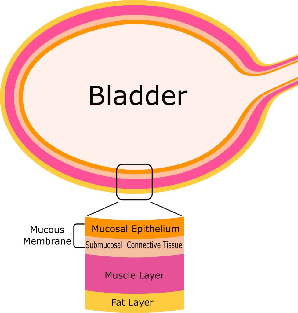
Fig. 1 Bladder layer structure The bladder is composed of a mucous membrane, a muscle layer, and a fat layer in order from the inside to the outside. The mucous membrane is further divided into the mucosal epithelium and submucosal connective tissue.
The bladder is an organ that temporarily retains urine produced by the kidneys and then releases it from the body. It consists of three layers. From the inside to the outside they are: a mucous membrane, a muscle layer, and a fat layer (Fig. 1). The mucous membrane is further classified into the mucosal epithelium and submucosal connective tissue. The number of newly diagnosed cases of bladder cancer in Japan is estimated to be 21,000 per year (from projected cancer statistics in 2019, based on the cancer registry and statistics of the National Cancer Center).
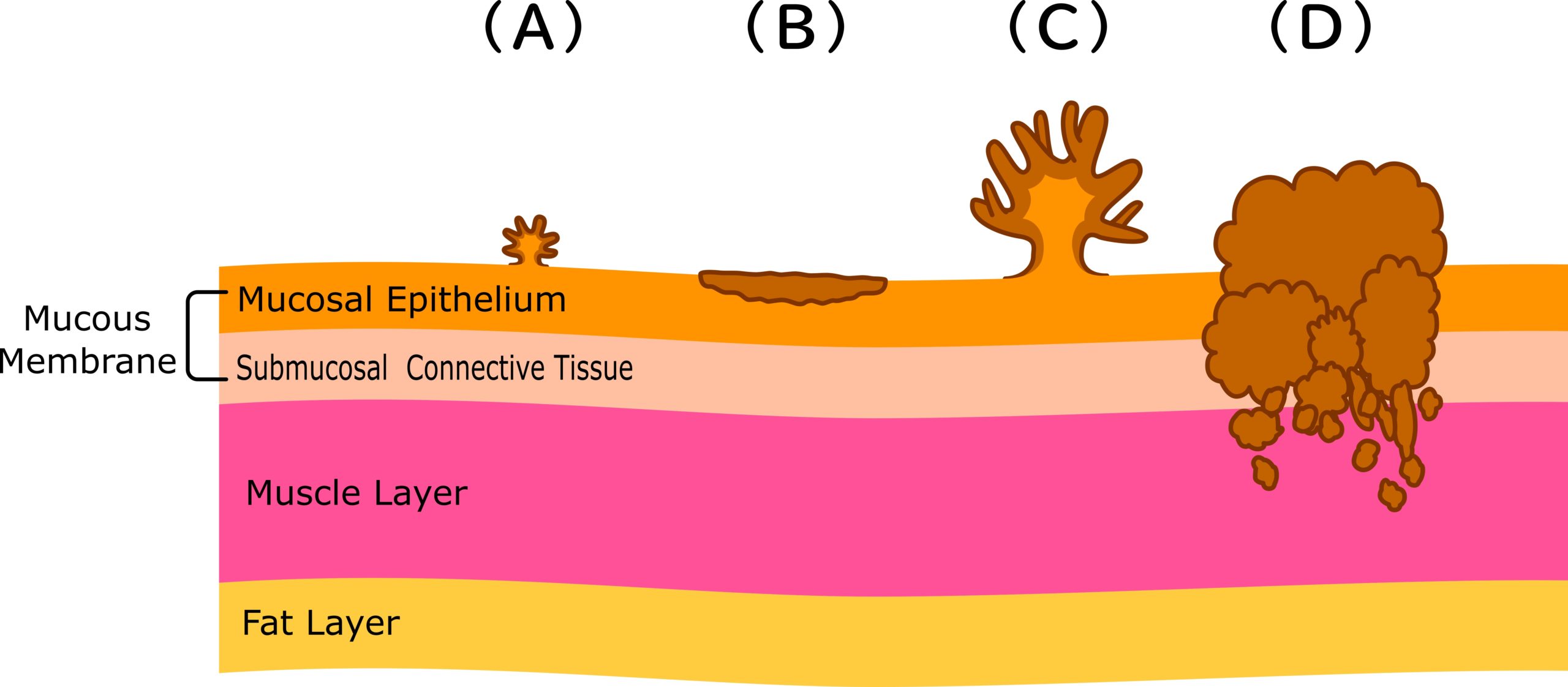
About 90% of bladder cancers occur in the mucous membrane. Cancer that remains in the mucous membrane and does not invade the muscle layer is called non-muscle-invasive bladder cancer (Fig. 2 A, B, C) and cancer that spreads to the muscle layer is called muscle invasive bladder cancer (Fig. 2 D).
Non-muscle-invasive bladder cancers include papillary (clinical) cancer that looks like sea anemones and protrudes into the lumen (Fig. 2 A, C), and a flattened intraepithelial cancer (Fig. 2 B).
Fig. 2 Image of bladder cancer Bladder cancers are categorized into two types: non-muscle-invasive bladder cancers that remain in the mucous membrane and do not invade the muscle layer (A, B C); and muscle-invasive bladder cancers that spread to the muscle layer (D). There are also small cancers (A) that are difficult to detect with a normal cystoscope, and flat cancers (cancers in the epithelium) that are difficult to distinguish from normal tissues (B).
The most common symptom of suspected bladder cancer is asymptomatic hematuria without pain. It includes not only hematuria that can be seen with the naked eye, but also hematuria that can be seen under the microscope. In addition to asymptomatic hematuria, frequent urination, a residual urinary sensation after urination, and pain during urination may occur. When asymptomatic hematuria exists or if bladder inflammation does not improve, an endoscopic examination is performed.
When bladder cancer is suspected, a cystoscopy and a urine cytology test are performed. Cystoscopy is a procedure in which a cystoscope (a thin tube with a camera on the tip) is inserted into the bladder from the urethra to observe inside the bladder, and ascertain the location, size, number, and shape of the cancerous cells. A urine cytology test is a test to check for the presence of cancer cells in the urine.
Depending on the results of the initial examination, an ultrasound examination (echo), computed tomography (CT), nuclear magnetic resonance examination (MRI), chest X-ray examination, or bone scintigraphy, etc. may be performed. For a definitive diagnosis of bladder cancer, collecting a bladder tissue sample for pathological exam is required.
(Supervised by Dr. Yoshihiko Hirao, Director Emeritus, Osaka Gyoumeikan Hospital, Professor Emeritus, Nara Medical University)